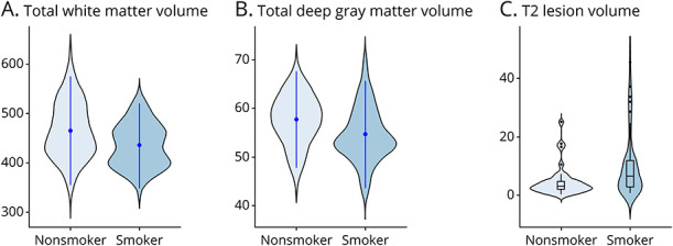
Figure 2. Distribution Plots of MRI Measurements at the 10-Year Follow-up Visit in Nonsmokers and Smokers.

The width of the shaded area represents the proportion of observations for (A) total white matter volume (mL) and (B) total deep gray matter volume (mL) (point range represents the mean and SD), and (C) T2 lesion volume (mL) (box plots represent the median and IQR, with the whiskers representing the distribution of observations within x1.5 of the IQR). IQR = interquartile range.
