Figure 5.
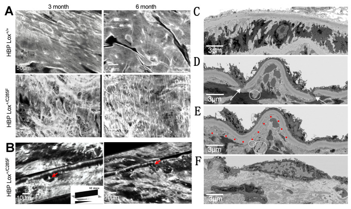
Increased fenestrations in Lox+/C285F elastic lamellae. (A), En-face two-photon imaging shows smooth and regular IEL in Lox+/+ (top) while Lox+/C285F are irregular and less tightly woven with numerous obvious fenestrations (bottom) See Supplemental Figure S1 for images of the second lamella). Representative images, n = 3. (B), Still images demonstrate fenestrae in 3D by crossing XY (10 μm) and XZ (5 μm) planes of the images in Lox+/C285F mice (see also Supplemental Videos S1–S6). (C–F), Still images from FIB-SEM show an intact IEL in Lox+/+ (C). In contrast, Lox+/C285F mice exhibit pathology ranging from significant disruptions/holes (arrows), disconnected “floating” segments (circled), and even sheared (red dotted line) stretches (D,E) to a severe, completely disorganized IEL (F) (See 3D reconstructed movies in Supplemental Videos S7–S9).

