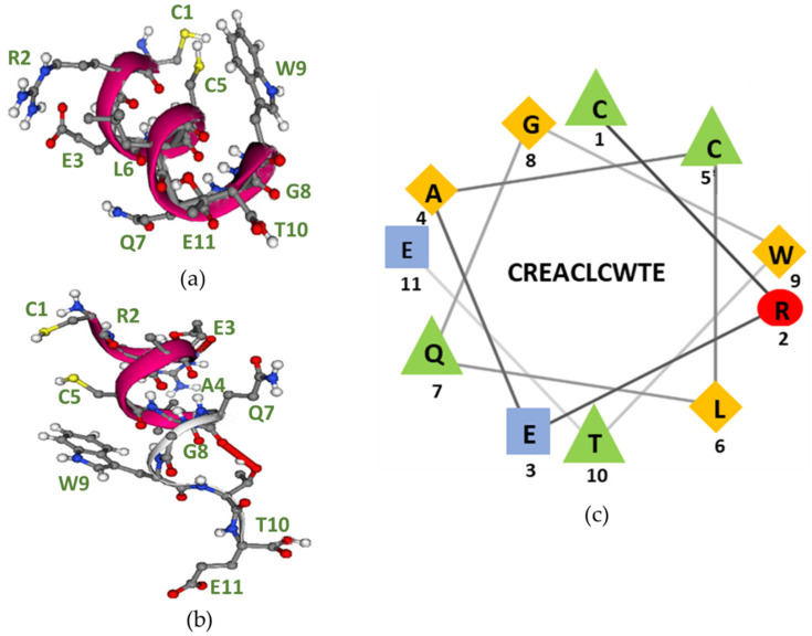
Figure 7.
Structural epitope analysis. (a) The three-dimensional structure of the CREACLQGWTE sequence obtained from PEP FOLD SERVER. The peptide folds into an α-helix structure in which protruding amino acid residues are seen. On the right side of the structure, the side-chain residues of Trp9, Cys1, and Cys5 protrude. On the left side of the structure, the side-chain residues of Arg2, Glu3, Leu6, Gln7, and the terminal Thr10 protrude. (b) The three-dimensional structure of the CREACLQGWTE peptide. Yellow balls = sulphur atoms, red balls = oxygen atoms, grey balls = carbon atoms, white balls = hydrogen atoms, and blue balls = nitrogen atoms. (c) Helical wheel representation of the CREACLQGWTE peptide. The residues in the epitope are presented using one-letter codes. The image was generated using the NetWheels application (http://lbqp.unb.br/NetWheels, accessed on 14 May 2022). The following colours represent amino acid functions: red, polar/basic; blue, polar/acid; green, polar/uncharged; yellow, nonpolar.

