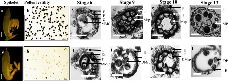
Figure 2.
The phenotype of S1 mutant. The spikelet of WT (a) and S1 mutant (g). The pollen fertility of WT (b) and the S1 mutant (h) by iodine staining magnified 20×. Transverse section analysis of the anther development in WT (c–f) and the S1 mutant (i–l). Locules from the anther section of WT and at stage 6 (the microspore mother cells stage), stage 9 (the young microspore stage), Stage 10 (the vacuolated pollen stage), and stage 13 (the mature pollen stage). E, epidermis; En, endothecium; T, tapetum; MMC, microspore mother cells; Msp, microspores; MP, mature pollen; DMsp, degenerated microspores; DP, degenerated pollen. Scale bar: 50 μm.

