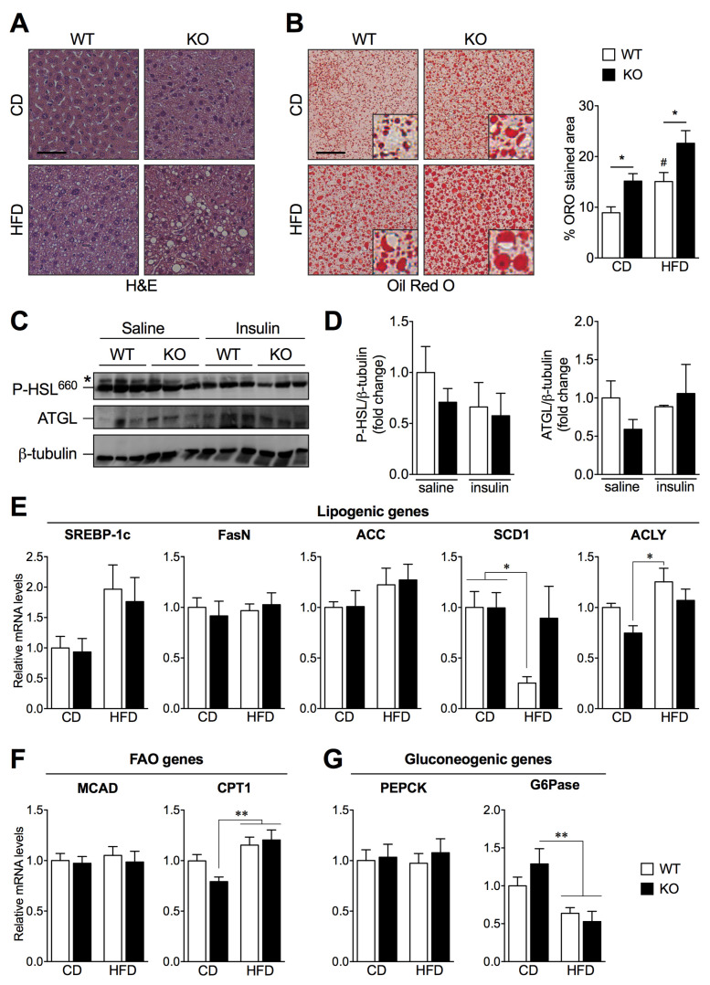
Figure 3.
Loss of SIRT2 promotes hepatic steatosis and alters the expression of key genes involved in de novo lipogenesis, fatty acid oxidation, and gluconeogenesis. (A) Representative images of H&E-stained and (B) ORO-stained liver sections of WT and SIRT2-KO mice fed a CD or a HFD, n = 5 per group. ORO stained area (%) was quantified using ImageJ software (scale bar = 50 μm). (C) Representative Western blots of adipose tissue lysates (50 μg) showing P-HSL and ATGL expression levels of WT and SIRT2-KO mice fed a HFD, upon saline or insulin stimulation (i.p. injection 2 U/kg). (D) Levels of P-HSL and ATGL were normalized to β-tubulin in the same lane. All P-HSL/β-tubulin and ATGL/β-tubulin ratios were normalized to the value from WT mice injected with saline. Each lane corresponds to a distinct animal. n = 3 per group. (E–G) RT-qPCR analysis of lipogenic (SREBP-1c, FasN, ACC, SCD1, and ACLY), fatty acid oxidation (MCAD, CPT1), and gluconeogenic (PEPCK and G6Pase) genes in livers of WT and SIRT2-KO mice fed a CD or a HFD. Results were normalized to reference gene Ywhaz and represented as mean ± SEM; n = 5–6 per group; # p < 0.05 compared with CD-fed WT mice; * p < 0.05, ** p < 0.01.

