
Figure 4.
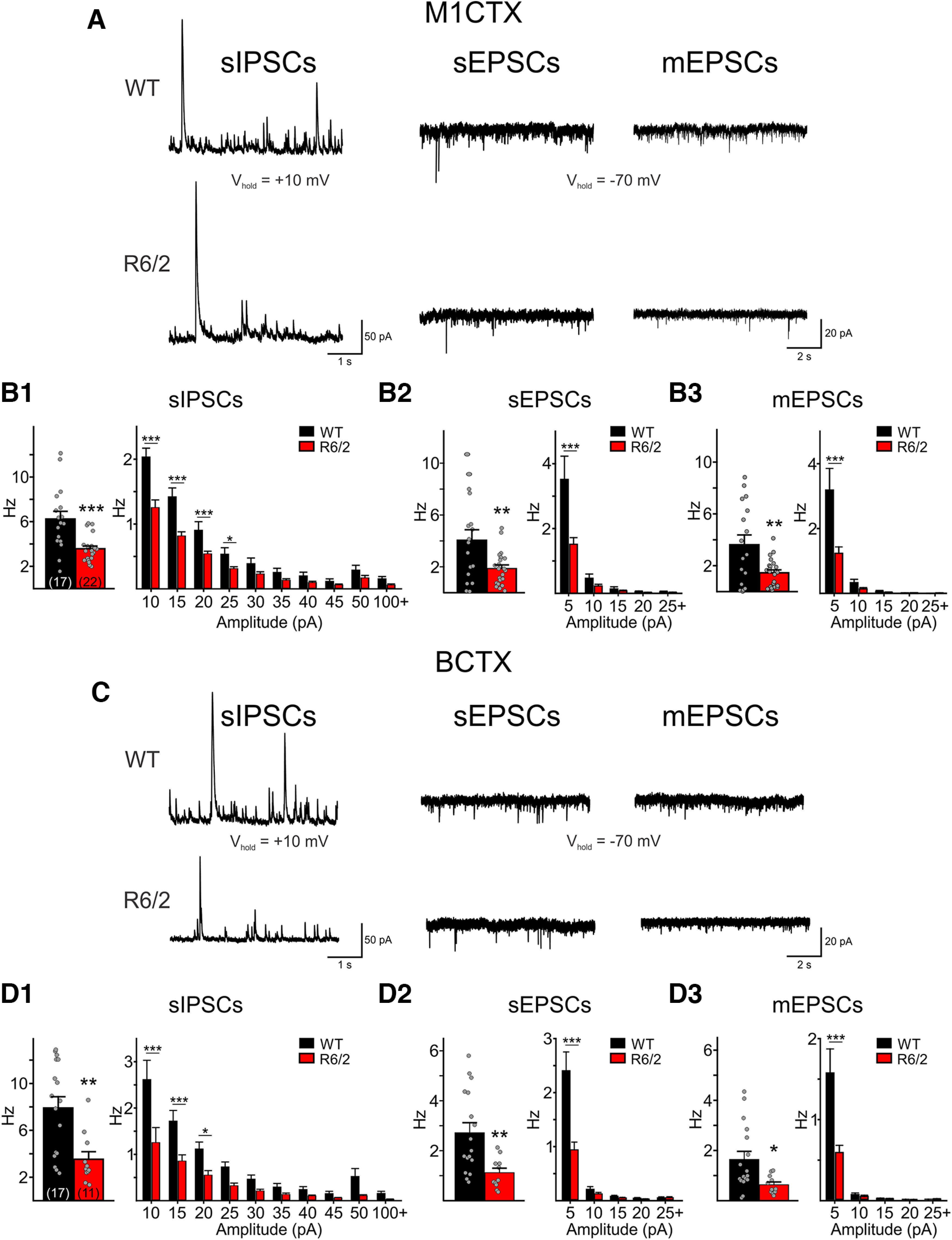
A, Sample raw traces of sIPSCs, sEPSCs, and mEPSCs recorded in M1CTX CPNs from a WT and a symptomatic R6/2 mouse. Summary of average frequencies of sIPSCs (B1), sEPSCs (B2), and mEPSCs (B3) in all recorded M1CTX CPNs (left). The number of recorded cells is shown in parentheses. Summary of average amplitude-frequency distribution plots (5-pA bins) are shown on the right. C, Sample raw traces of sIPSCs, sEPSCs, and mEPSCs recorded in a BCTX CPN from a WT and a symptomatic R6/2 mouse. Summary of average frequencies of sIPSCs (D1), sEPSCs (D2), and mEPSCs (D3) in all recorded BCTX CPNs (left). Average amplitude-frequency distribution plots (5-pA bins) are shown on the right. Significant differences between genotypes were determined using Student’s t tests and two-way repeated measures ANOVAs followed by Bonferroni post hoc tests; *p < 0.05, **p < 0.01, and ***p < 0.001.