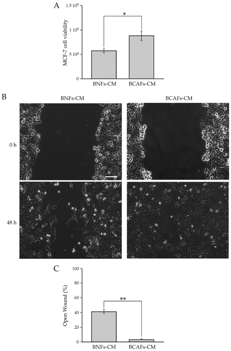
Figure 2.
(A) Influence of conditioned media from BNFs (BNFs-CM) and BCAFs (BCAFs-CM) on MCF-7 cell viability. Data are means of at least three independent experiments ± S.E. * p < 0.05. (B,C) Influence of conditioned media from BNFs (BNFs-CM) and BCAFs (BCAFs-CM) on MCF-7 cell migration. (B) Wound-Healing Assay performed on MCF-7 cells treated with fibroblast-conditioned media. Representative images of three independent experiments show the same fields with scratching at 0 and 48 h after wounding. Scale bar 100 µm. Magnification ×10. (C) Migratory capability quantification of MCF-7 cells. Wound widths were measured at 0 and 48 h after wounding. Data are expressed as percentage of fold decrease in open wound area compared with control (0 h) set as 100% and are reported as mean of three independent experiments ± S.E. ** p < 0.0001.

