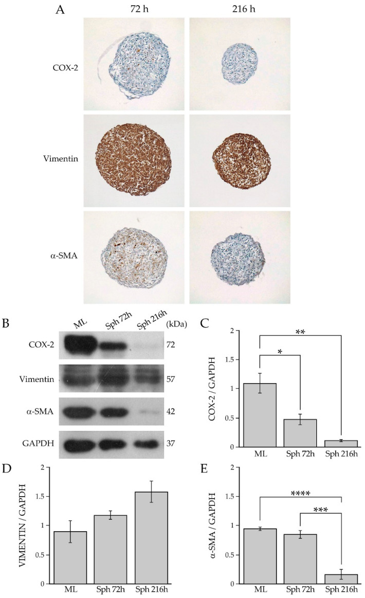
Figure 5.
(A) Immunohistochemical analysis of COX-2, vimentin and α-SMA in BCAF spheroids collected at 72 h and 26 h. Magnification ×10. (B) Western blotting analysis of COX-2, vimentin, and α-SMA in protein extracts of BCAF monolayers (ML) and BCAF spheroids collected at 72 h (SPH 72 h) and 216 h (SPH 216 h) of 3D cell culture on agar. GAPDH was used as the loading control. Representative images of three independent experiments are shown. Densitometric analyses of (C) COX-2, (D) vimentin and (E) α-SMA protein levels. Data are reported as means of three independent experiments ± S.E. * p < 0.05, ** p < 0.005, *** p < 0.001, **** p < 0.0005.

