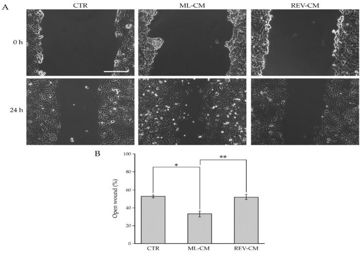
Figure 8.
Influence of BCAF monolayers and reverted BCAFs on the migratory capability of BC cells. (A) Wound-Healing Assay of MCF-7 cell line grown for 24 h with the conditioned media from BCAF monolayers (ML CM) and reverted BCAFs (REV CM). Standard cell culture medium was used as control. The representative images of three independent experiments show the same fields with scratching at 0 and 24 h after wounding. Scale bar 200 µm. Magnification ×10. (B) Migratory capability quantification of MCF-7 cells. Wound widths were measured at 0 and 24 h after wounding. Data are expressed as percentage of fold decrease in open wound area compared with control (0 h) set as 100% and are reported as mean of three independent experiments ± S.E. * p < 0.0005, ** p < 0.0001.

