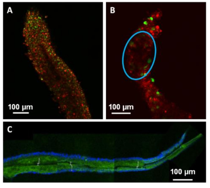
Figure 6.
Confocal microscope images showing laser-induced modifications in three different types of microfibres. (A) Sharp channel inside a dehydrated microfibre after spending 41 min on a sample holder. (B) Attenuation (blue circle) of signal density inside a hydrated microfibre after 9 min spent on a sample holder. After being marked with Live/Dead® ki, A and B colors were modified to highlight laser interaction effects. (C) Sharp channel inside a PFA-fixed microfibre, green: collagen-Fluorescein-5-isothiocyanate (FITC), blue: DAPI staining.

