Figure 2.
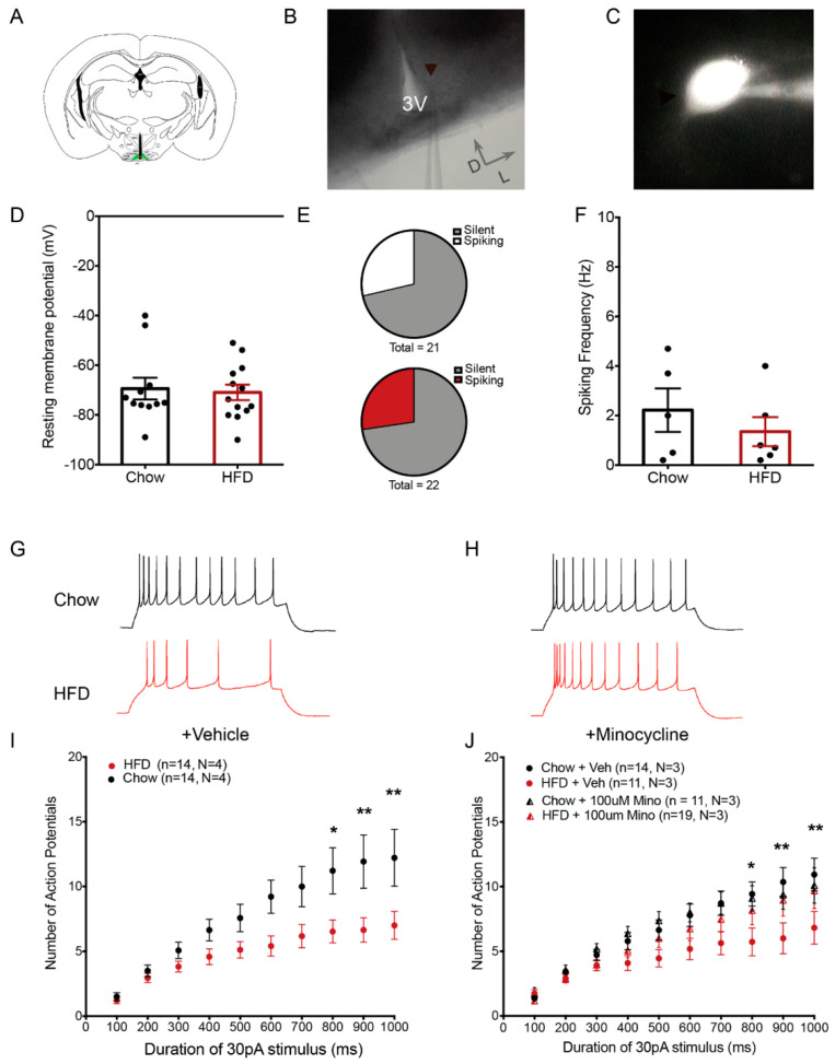
Minocycline restores intrinsic excitability of POMC neurons in HFD-fed mice. (A) Schematic diagram showing a coronal hypothalamic slice. The arcuate nucleus (ARC), where GFP-expressing POMC neurons were visualized and recorded from, is highlighted in green. (B) 4X DIC image showing the ARC region and recording pipette attached to a recorded POMC neuron. (C) Representative fluorescent image of a GFP-expressing POMC neuron with attached recording pipette filled with internal solution containing fluorophore Alexa-594. (D) Resting membrane potential of recorded POMC neurons (Chow: −69.38 ± 4.3 mV, n = 11, N = 3; HFD: −70.92 ± 3.1 mV, n = 13, N = 3). (E) Proportion of recorded POMC neurons showing spontaneous activity (upper: Chow (6 out of 21 neurons), lower: HFD (6 out of 22 neurons)). (F) Frequency of action potentials in spontaneously firing POMC neurons (Chow: 2.220 ± 0.87 Hz, n = 6, N = 5; HFD: 1.35 ± 0.58 Hz, n = 6, N = 5). (G,H) Representative traces of POMC neuron responses after stimulation by 1000 ms 30 pA current step in brain slices from chow and HFD-fed mice treated with vehicle (G) or minocycline (100µM) (H). (I,J) Population data of evoked responses of POMC neurons to variable duration of stimulus in brain slices from chow and HFD-fed mice incubated with vehicle (I) or minocycline (J). Lowercase (n) indicates number of neurons and uppercase (N) number of animals. * p < 0.05; ** p < 0.01.

