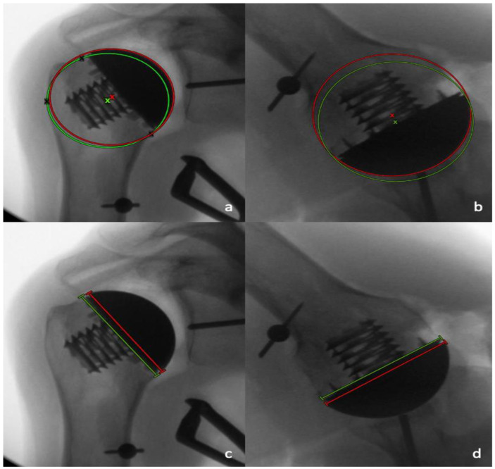
Figure 5.
A best-fit anatomic circle (green circle) with its center of rotation (COR) (green x), and a best-fit implant circle (red circle) with its COR (red x) are placed on the AP (a) and axillary (b) views to determine the differences in the positioning of the COR. The length of the resection plane (green line) and the prosthetic humeral head (red line) were compared on the AP radiographs (c) and the axillary radiographs (d).

