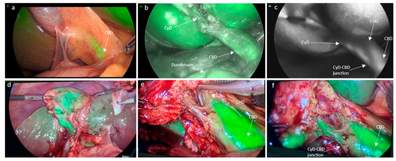
Figure 2.
Intravenous ICG injection. (a) Initial view of ICG-cholangiography prior to dissection; (b) visualization of cystic duct after dissection; (c) view in ‘SPY’ mode; (d) clear delineation of the gallbladder; and (e,f) cystic and common bile duct with minimal liver fluorescence (CyD: cystic duct, CyA: cystic artery, CBD: common bile duct, CHD: common hepatic duct).

