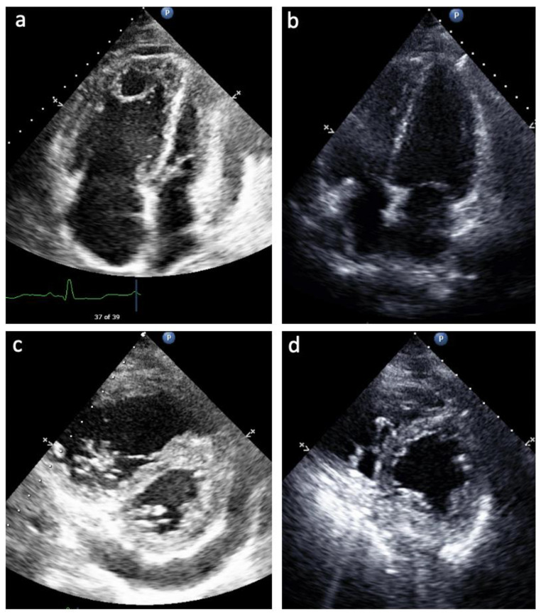
Figure 1.
Echocardiogram from Patient 3 prior to (a,b) and after (c,d) PAH medication optimization. (a) Apical 4 Chamber view of enlarged RA and enlarged and hypertrophied RV with small and underfilled LV, LA; (c) Apical 4 Chamber view of normalized RA and RV size and function on PAH therapy; (b) Parasternal short axis view of severe systolic septal flattening, RV enlargement and hypertrophy, and pericardial effusion; (d) Parasternal short axis view of resolution of systolic septal flattening and pericardial effusion with smaller RV size.

