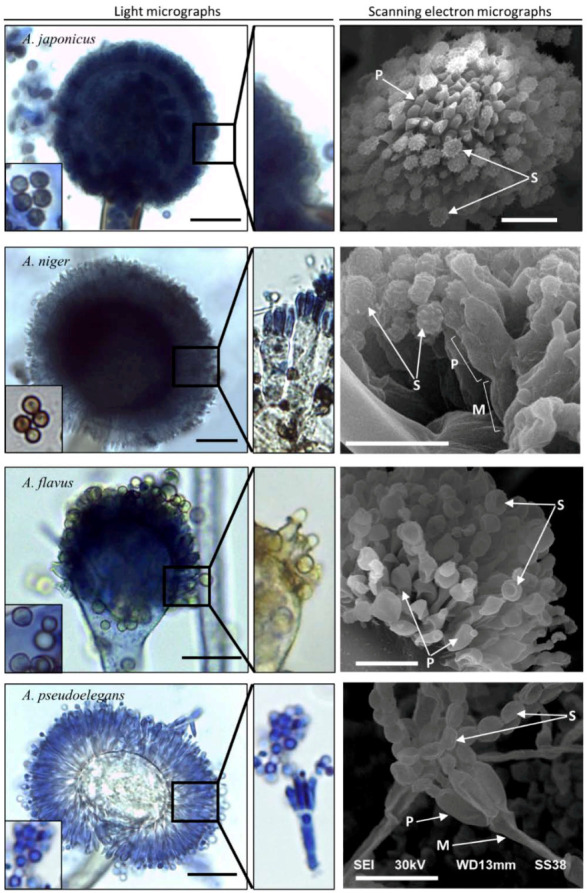
Figure 2.

Light and scanning electron microscopic characterization of Aspergillus spp. Spores (S) are represented in the small bottom left panel in each photo. Phialides (P) and seriation types are shown in each side panel under higher magnification power. Metulae (M) is indicated in biseriate fungal heads. The scale bar is 10 µm in both light and electron micrographs.
