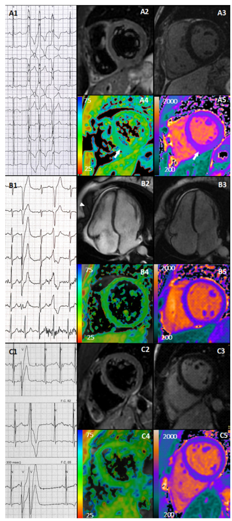
Figure 1.
Newly detected abnormal findings. Case 1. Athletes presented with exercise-induced NSVT with LBBB morphology, inferior axis and transition in V3 (A1), negative T2-weighted (A2), absence of LGE (A3), increased T2 mapping (A4), and native myocardial T1 mapping (A5) at the mid-inferior wall and inferior septum (white arrows). Case 2. Athlete presented with exercise-induced polymorphic PVCs with LBBB morphology with transition in V6 (first one) and in V4 (second one) (B1), cine CMR with mild pericardial effusion (arrows) (B2), negative LGE (B3), normal T2 mapping (B4), and T1 myocardial native mapping (B5). Case 3. Athlete presented with exercise-induced polymorphic PVCs, often R on T, and a couplet (C1) and negative T2-weigthed sequence (C2), negative LGE (C3), normal T2 mapping (C4), and native myocardial T1 mapping (C5).

