. 2022 Jun 8;11:e77115. doi: 10.7554/eLife.77115

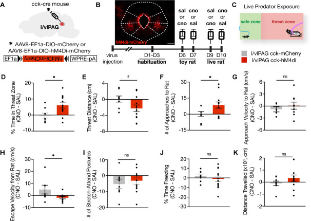
Figure 7. Lateral/ventrolateral periaqueductal gray-cholecystokinin-expressing (l/vlPAG-CCK) inhibition increases time spent near a live predator and reduces escape vigor without altering freezing.

(A) Viral strategy for bilateral expression of inhibitory designer receptor hM4Di-mCherry or mCherry in CCK cells in the l/vlPAG. (B) Top-left: expression of hM4Di-mCherry in l/vlPAG-CCK cells. Scale bar, 200 μm. Bottom: timeline for DREADD experiments. Saline or clozapine-N-oxide (CNO, 10 mg/kg) occurred 40 min prior to exposure. (C) Live predator exposure schematic. Mice were placed in the presence of an awake rat restrained to one end of a chamber. Each exposure lasted 10 min. (D–K) Chemogenetic inhibition of l/vlPAG CCK cells increased time spent in threat zone (D, unpaired t-test, *p=0.0199), increased number of approaches toward the rat (F, unpaired t-test, *p=0.0496), and reduced escape velocity from the rat (H, unpaired t-test, *p=0.0431). Threat distance trended toward significance with CCK inhibition (E, unpaired t-test, #p=0.069). Approach velocity (G), stretch-attend postures (I), freezing (J), and distance traveled (K) were unaltered with inhibition (D–F, I–K: mCherry, n = 8; hM4Di, n = 12; G: mCherry, n = 5, hM4Di, n = 9; H: mCherry, n = 7, hM4Di, n = 11). Errorbars: mean ± SEM.

Figure 7.

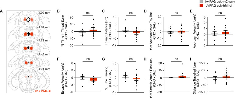
Figure 7—figure supplement 1. Chemogenetic of lateral/ventrolateral periaqueductal gray-cholecystokinin-expressing (l/vlPAG-CCK) population using DREADDs during exposure to a toy rat.

Figure 7—figure supplement 1.

(A) Bilateral cre-dependent HM4Di expression in l/vlPAG of Cck-ires-cre mice. (B–I) Chemogenetic inhibition of l/vlPAG-CCK cells during exposure to toy rat (mCherry, n = 8; hM4Di, n = 12; unpaired t-test). Errorbars: mean ± SEM.
Figure 7—figure supplement 2. Inhibition of lateral/ventrolateral periaqueductal gray-cholecystokinin-expressing (l/vlPAG-CCK) neurons does not alter pain response latency or acquisition of learned fear.
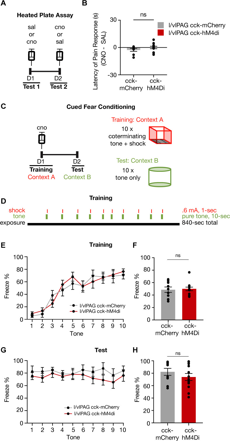
Figure 7—figure supplement 2.

(A) Timeline of heated plate assay with chemogenetic inhibition of l/vlPAG-CCK cells. (B) Inhibition of l/vlPAG-CCK cells does not alter latency of pain response (clozapine-N-oxide [CNO] minus saline; mCherry, n = 8; hM4Di, n = 12; unpaired t-test). Mean ± SEM. (C) Timeline of cued fear conditioning across 2 days (training and test) with l/vlPAG-CCK chemogenetic inhibition during training. Training occurred in context A, which consisted of metal bar flooring, bare gray walls, warm-colored lighting, and cleaned with 70% ethanol. Training included tone-shock pairings. Test occurred in context B, which consisted of rounded white walls, gray smooth flooring, blue-colored lighting, and cleaned with Strike-Bac. Both contexts A and B were illuminated to 40 lux. Test included tone presentations only. (D) Schedule of tone and shock presentations during training. Trial was 14 min in total, with 10 pairs of co-terminating 10 s tones and 1 s shocks. The tone was a 70 dB pure-tone, and all shocks were 0.6 mA. (E) Mean freezing of CCK-mCherry and CCK-hM4Di mice during 10 s tone presentations during training (mCherry, n = 9; hM4Di, n = 12). (F) No difference in freezing during training between groups (mCherry, n = 9; hM4Di, n = 12; unpaired t-test, p=0.799). (G) Same as (E) but during test (mCherry, n = 9; hM4Di, n = 12). (H) Same as (F) but during test (mCherry, n = 9; hM4Di, n = 12; unpaired t-test, p=0.328). Errorbars: mean ± SEM.