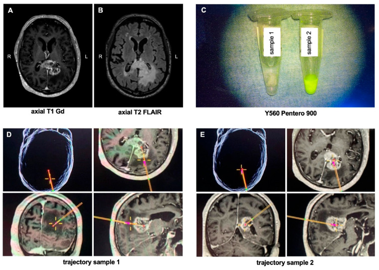
Figure 3.
Illustrative case. (A) Axial Gadolinium-enhanced T1-weighted and (B) T2 FLAIR MRI of a 72-year-old patient who presented with a left-sided contrast-enhancing lesion with perifocal edema of the splenium of the corpus callosum. The patient underwent a VarioGuide® biopsy in which a total of 8 samples were taken, of which two are exemplary shown: (C) image of the specimen under the Y560 filter of the Pentero 900 microscope; (D,E) intraoperative trajectory of the two obtained samples; sample 1 was taken from the infiltration zone of the tumor without contrast-enhancement, whereas sample 2 was obtained from tumor core. Abbreviations: Gd = gadolinium; L = left; R = right.

