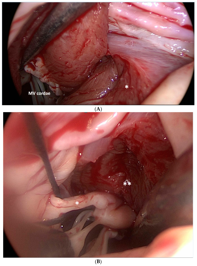
Figure 6.
Surgical myectomy (*) was performed starting at the nadir of the right coronary sinus, and extended apically to achieve the exposure of the papillary muscles. MV: mitral valve (A). Surgical view of the diseased mitral valve secondary cordae (*) and papillary muscles (**) after myectomy. It is noteworthy that the bases of the papillary muscles are now visible (B).

