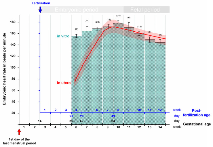
Figure 5.
This graph depicts the changes in the contraction rate of human embryonic and fetal hearts during the first trimester of pregnancy as recorded (1) in vitro on isolated heart specimens obtained from aborted embryos and fetuses (data from [147]; mean values ± S.E.; number of cases in parentheses); and (2) in utero by transvaginal ultrasonography (data from [30]). Note that, during the first 3 weeks of heart activity, the in vitro values are markedly higher than those measured in utero. From the seventh post-fertilization week (ninth gestational week) onward, however, in vitro and in utero values do not differ markedly from each other.

