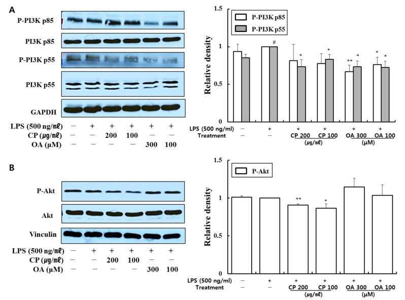
Figure 4.
Effects of CP and OA treatments on the cellular protein levels of the PI3K/Akt pathway in LPS-stimulated LA4 cells. LA4 cells were stimulated with LPS for 1 h and then incubated with CP or OA treatments for 24 h. The proteins of (A) P-PI3K p85 (Tyr458), P-PI3K p55 (Tyr199), total PI3K p85, total PI3K p55, and (B) P-Akt (Ser473), total Akt were measured by Western blot. The images shown are representative, and GAPDH and vinculin were used as internal controls. The densities of the bands were assessed by Image J. The data are presented as the mean ± SEM; # p < 0.05 vs. the normal group; * p < 0.05 and ** p < 0.01 vs. the LPS-stimulated group. P-, phosphorylated.

