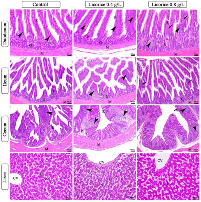
Figure 1.
Photomicrograph of duodenum, ileum, cecum, and liver of broiler chickens of control, 0.4 gm/L and 0.8 gm/L licorice acid groups. The panels of the duodenum, ileum, and cecum show simple columnar epithelium of lamina epithelial (E), lamina propria submucosa (P), mucosal glands (G), tunica muscularis (M), lymphatic tissue in both lamina epithelial and lamina propria (arrowheads), congested blood vessels of lamina propria (arrows). The panels of the liver show central vein (CV), hepatocytes (H), and blood sinusoids (S), which appear dilated and congested in the 0.8 gm/L licorice acid group. Stain H&E.

