
Figure 8.
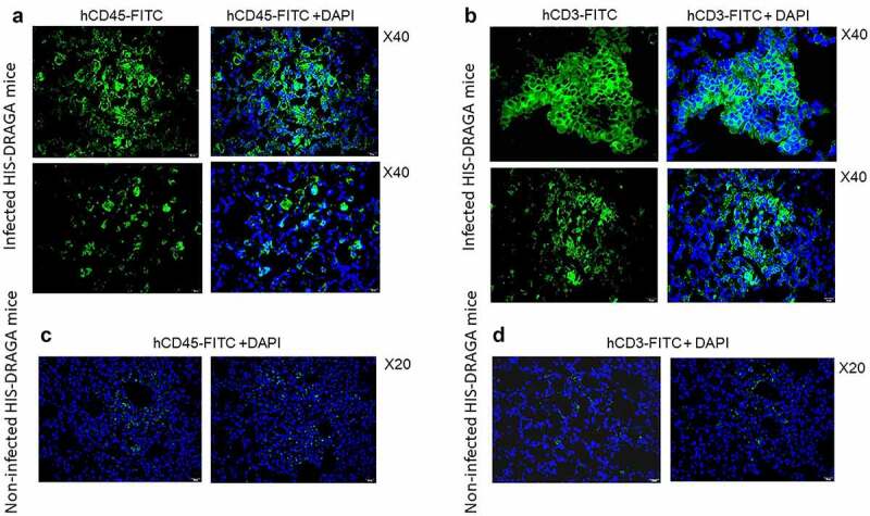
Infiltrating human lymphocytes in the lungs of SARS-CoV-2 infected HIS-DRAGA mice.
Lung sections from HIS-DRAGA female mice #F4 (upper panels in a & b) and #F3 (lower panels in a & b) that had recovered from SARS-CoV-2 infection(103 pfu/mouse) by 25 dpi stained for hCD45 and hCD3. Of note, infiltrating hCD45+ lymphocytes and hCD3+ T cells in the infected mice were more abundant and organized in clusters than those in non-infected mice that were dispersed through parenchyma (panels & d).