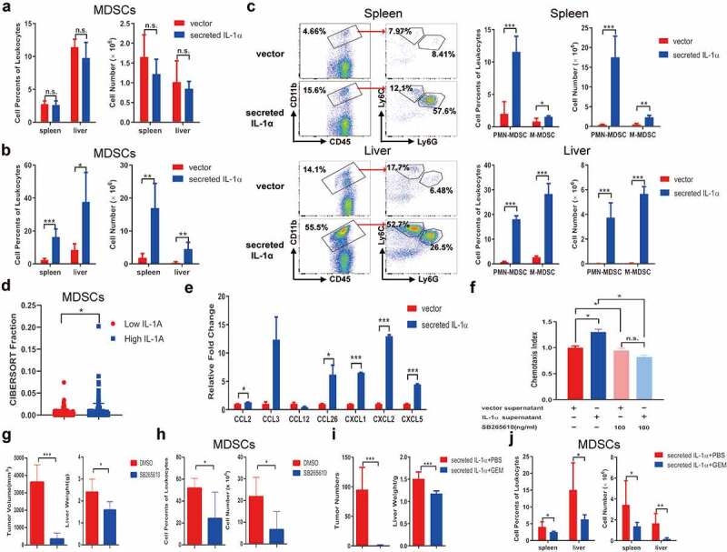
Figure 3.

Tumoral-secreted IL-1α increases MDSCs in vivo.
Splenocytes and intrahepatic leukocytes were isolated from orthotopic HCC mice (n = 5–7 each group). CD11b+Gr1+ MDSCs in spleen and liver of the tumor-bearing mice expressing secreted IL-1α or vector control were analyzed by flow cytometry 1 week (a) or 3 weeks (b) after tumor injection. (c) The percentages and total numbers of PMN-MDSCs and M-MDSCs were analyzed by flow cytometry. (d) CIBERSORT analysis of the relationship between IL1A expression and MDSCs infiltration in HCC patients. (e) Multiple chemokines and cytokines expressions were assessed by qPCR from tumor cells. (f) MDSCs were preincubated with CXCR2 inhibitor SB265610 for 30 min to block CXCR2 interaction with its ligands. Transwell assay was performed to detect the chemotaxis ability. (g) Mice were treated with SB265610 at a dose of 2 mg/kg mouse body weight every 3 days (n = 5 each group). The tumor size and weight were measured. (h) The percentage and total number of tumoral MDSCs infiltration was detected by flow cytometry. (i) Tumor-bearing mice expressing secreted IL-1α were injected with PBS or GEM (n = 5–7 each group). The numbers of tumor nodules and liver weight were shown. (j) The depletion of MDSCs was confirmed by flow cytometry. The data shown are the representative of three experiments. Data are presented as means ± SD. * p < .05, ** p < .01, *** p < .001.
