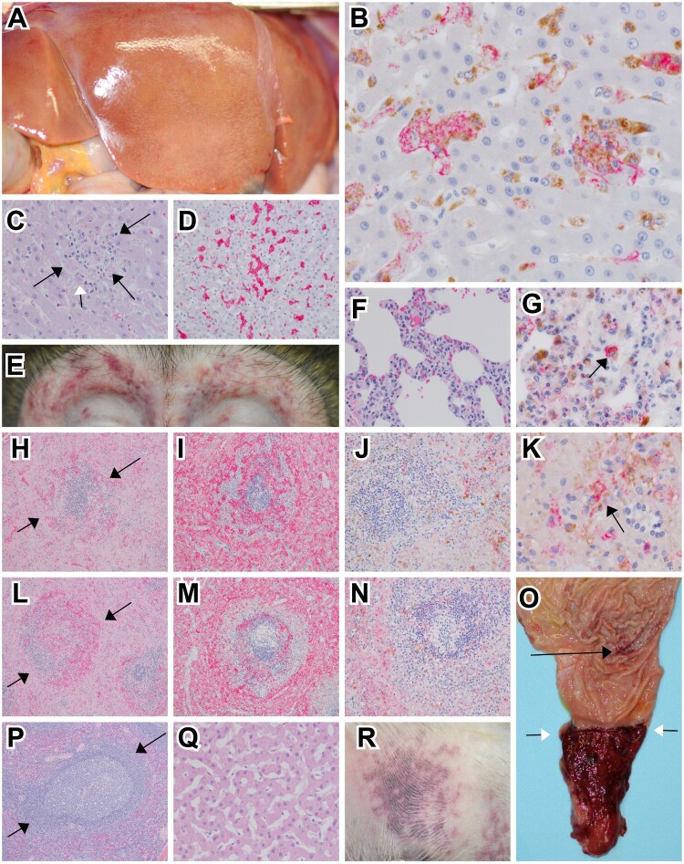
Figure 3.
Representative gross and histologic lesions of SUDV-infected macaques. (A) Diffuse hepatic pallor indicative of hepatitis in cynomolgus macaque (RHES-1). (B) Immunohistochemistry (IHC) double labelling for macrophage (CD68-brown) and SUDV VP40 (red) in the liver of rhesus macaque (RHES-6) 40x, normal hepatic architecture was disrupted with expanded sinusoids that were occupied with clusters of macrophages infected with SUDV. (C) Hematoxylin and eosin (H&E) staining of the liver in rhesus macaque (RHES-6) 40x, normal hepatic architecture was disrupted with expanded sinusoids with mononuclear inflammation and eosinophilic cellular debris (black arrows), rarely eosinophilic intracytoplasmic inclusion bodies were noted (white arrow). (D) IHC for fibrin (red) in rhesus macaque (RHES-6) 20x, intra-sinusoidual fibrin deposition. (E) Bilateral periorbital petechial rash in rhesus macaque (RHES-2). (F) H&E staining of lung in cynomolgus macaque (CYNO-9) 40x, expansion of alveolar septa with mononuclear inflammatory cells and minimal extravasation of erythrocytes within alveoli. (G) IHC double labelling for macrophage (CD68-brown) and SUDV VP40 (red) in the lung of cynomolgus macaque (CYNO-9) 60x, alveolar macrophages were infected with SUDV (black arrow). (H) H&E staining of spleen in rhesus macaque (RHES-6) 10x, loss of normal white pulp architecture with extensive lymphocytolysis, hemorrhage and fibrin deposition (black arrows). (I) IHC labelling for fibrin (red) in the spleen of rhesus macaque (RHES-6) 10x, extensive fibrin deposition in red and white pulp. (J) IHC double labelling for macrophage (CD68-brown) and SUDV VP40 (red) in the spleen of rhesus macaque (RHES-6) 20x, macrophages infected with SUDV were sparsely present in the red and white pulp. (K) Higher magnification of IHC double labelling for macrophage (CD68-brown) and SUDV VP40 (red) in the spleen of rhesus macaque (RHES-6) 60x, macrophages infected with SUDV (black arrow). (L) H&E staining of spleen in cynomolgus macaque (CYNO-9) 10x, loss of normal white pulp architecture with extensive lymphocytolysis, hemorrhage and fibrin deposition (black arrows). (M) IHC labelling for fibrin (red) in the spleen of cynomolgus macaque (CYNO-9) 10x, extensive fibrin deposition in red and white pulp. (N) IHC double labelling for macrophage (CD68-brown) and SUDV VP40 (red) in the spleen of cynomolgus macaque (CYNO-9) 20x, macrophages infected with SUDV were sparsely present in the red and white pulp. (O) Mucosal surface of the pyloric region of the stomach and aboral duodenum in rhesus macaque (RHES-2), multifocal pinpoint ulcerations of the gastric mucosa (black arrow) and diffuse ulceration and hemorrhage of the aboral duodenum extending up to the pyloric sphincter (white arrows). (P) H&E staining of spleen in SUDV survivor rhesus macaque (RHES-7) 10x, no signification lesions. (Q) H&E staining of liver in SUDV survivor rhesus macaque (RHES-7) 10x, no signification lesions. (R) Petechial rash of the extremity in rhesus macaque (RHES-2).

