
Figure 5.
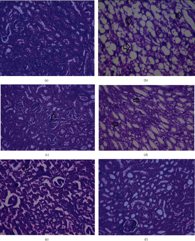
Histopathological photomicrographs of the kidney of diabetic obese rat treated with Malva neglecta extract at 40× magnification. Kidney of (a) normal rats. (b) Diseased control rats showing the necrosis of tubular cells. (c) Metformin treated animals showing the recovered epithelial and tubular cells. (d) MNME 250 mg/kg treated rats displaying the destroyed epithelial structures. (e) MNME 500 mg/kg treated rats showing the improved tubular and epithelial cell structure. (e) Rat treated with MNME 750 mg/kg exhibiting improved tubular and epithelial cell. Here, the arrow showed intact glomeruli, and the triangle showed slugged off epithelial cells and deranged tubular structure.