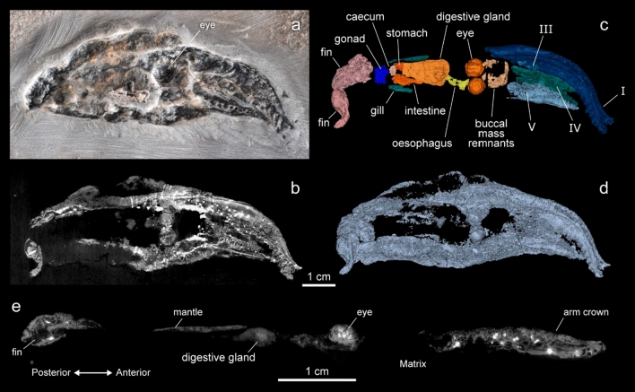
Figure 1.
Images and reconstruction of the V. rhodanica holotype (MNHN.B.74247) acquired using PPC-SRµCT (voxel size: 12.64 µm), at the ESRF (Grenoble, France). (a) Photograph (P. Loubry, CR2P) of the specimen showing the 3-D preservation of the mineralised soft tissue. (b) PPC-SRµCT slice showing the contrast in the grey-scale image used to segment the specimen. This contrast results from the density variation among the various mineralised tissues. (c) 3D representation showing the arm crown (arm pair I, III, IV, and V), as well as other presumed internal elements (d) 3D reconstruction of the whole specimen (e) Sagittal slice showing the profile view.

