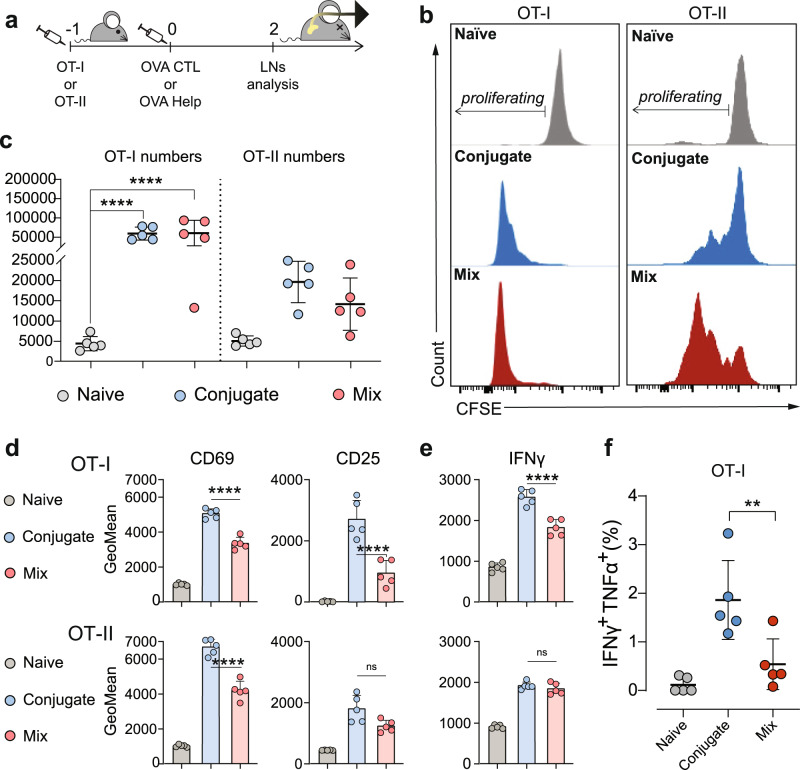
Fig. 3. OVA CTL and Help Lipid A-peptide conjugates promote enhanced activation of T cells upon in vivo injection.
a Mice (n = 5 per group) were adoptively transferred with CFSE labeled OT-I or OT-II cells 24 h before intradermally receiving 2 nmol of OVA CTL or Help CRX-527conjugates, or an equimolar mix of peptide and CRX-527. 48 h later, the inguinal lymph nodes were harvested for analysis of OT-I or OT-II T cell proliferation and activation. b Representative histograms of CFSE signal in labeled OT-I or OT-II cells. c Absolute count of total OT-I and OT-II cells in pooled inguinal lymph nodes. d Mean fluorescence intensity (GeoMean) of CD69 and CD25 T cell activation markers in OT-I (upper) or OT-II (lower) cells as detected by flow cytometry. e Mean fluorescence intensity of IFNγ cytokine in OT-I (upper) or OT-II (lower) as detected by flow cytometry. Statistical significance of the conjugates compared to the mix in both (d) and (e) was determined by one-way ANOVA followed by Sidak’s multiple comparison test; *****p < 0.0001. f Percentage of IFNγ/TNFα double producing OT-I cells. Statistical significance was determined by one-way ANOVA followed by Tukey’s multiple comparison test; **p < 0.01, all data are displayed as mean ± SD.

