Fig. 3.
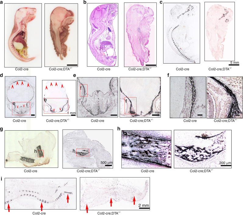
Ablation of Col2+ cells results in absence of the spine and disruption of endochondral skeletal development. a Total view of middle sagittal sections of Col2-cre and Col2-cre;DTA+/− mice at P0. b H&E staining of the middle sagittal sections of Col2-cre and Col2-cre;DTA+/− mice at P0. c Von Kossa staining of the middle sagittal sections of Col2-cre and Col2-cre;DTA+/− mice at P0. d Von Kossa staining of the skull with transection. Yellow arrow, severe bone loss in the back of the skull of Col2-cre;DTA+/− mice. Yellow star, nasal cavity area in Col2-cre and Col2-cre;DTA+/− mice. e Von Kossa staining of craniofacial bone showing loss of the nasal cavity in mutant mice. f Higher magnification of Von Kossa staining showing similar sponge-like bone structures in both wild-type and mutant mice. g Von Kossa staining of bone in the hindlimbs of wild-type and mutant mice. h Higher magnification of Von Kossa staining of the hindlimbs of wild-type and mutant mice. Note that a small piece of well-calcified sponge-like bone structure can be detected in mice with ablation of Col2+ cells. i Von Kossa staining of the middle sagittal section of the spine of wild-type and mutant mice. Red arrow, severe vertebral bone loss in Col2-cre;DTA+/− mice. n = 6 mice per condition; three independent experiments

