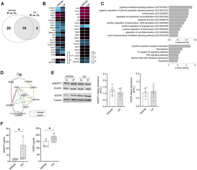
Fig. 4.
Identification and validation of the synergic mechanisms of action of CA. A Venn’s diagram of the differentially expressed proteins between the IP and CL brain hemispheres of vehicle- and CA-treated mice evaluated in the Olink proteomic array. B Heat maps of the differentially expressed proteins between the IP and CL brain hemispheres of mice treated with vehicle (left) or CA (right). C Biological significance analysis of the proteins modulated by CA. D Predicted molecular mechanism of action of CA on ischemic stroke obtained from the in silico generated mathematical models of stroke. E Representative Western blot images and quantification of brain levels of ANGPT1 and EGFR in vehicle- and CA-treated animals (n = 5/group). The ratio between IP and CL levels within each animal is depicted. F Quantification of ANGPT1 and EGFR levels in mouse plasma samples (n = 7/group). In all cases, mean ± SD is shown. * indicates p < 0.05 and **p < 0.01. Abbreviations: ANGPT1, angiopoetin-1; CCKAR, cholecystokinin receptor type A; CL, healthy contralateral brain hemisphere; EGFR, epidermal growth factor receptor; ELNE, neutrophil elastase; GNA11, G protein subunit alpha 11; HIF1A; hypoxia-inducible factor 1-alpha; IP, ipsilateral brain hemisphere; PK3CA, phosphatidylinositol-4,5-bisphosphate 3-kinase catalytic subunit alpha

