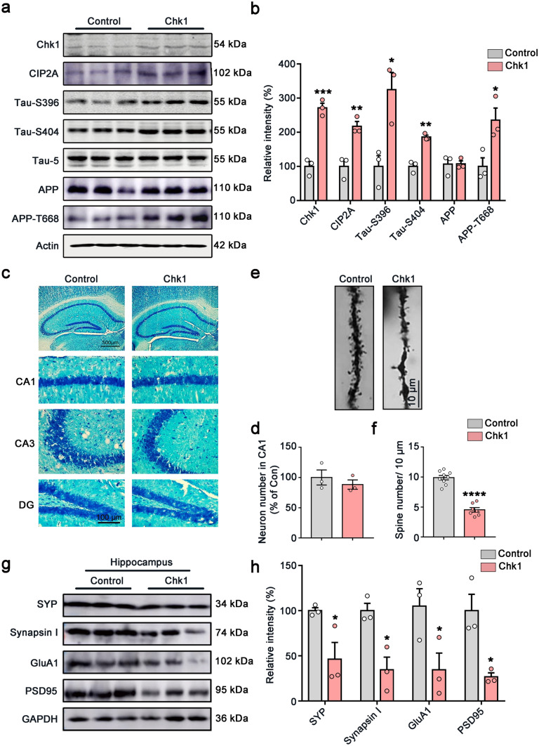
Fig. 4.
Overexpression of Chk1 in neurons induces CIP2A upregulation, tau/APP hyperphosphorylation, and synaptic impairments in mice. a Representative immunoblots of Chk1, CIP2A, Tau-S396, Tau-S404, Tau-5, APP, APP-T668, and β-actin in the hippocampus of the mice. Blots were from different gels with the same batch of samples electrophoresed. b The quantitative analysis of the protein level in a. Non-phosphorylated proteins such as Chk1, CIP2A, and APP were normalized to the β-actin levels; phosphorylated Tau-S396, Tau-S404, and APP-T668 were normalized to corresponding total tau (Tau-5) and APP levels respectively. c Representative images of Nissl staining of the mice brain slices. d The quantitative analysis of the neuron number in the CA1 region of c. The cell number was counted in brain slices from three mice in each group; one brain slice was counted for each mouse, and the neuron number in the CA1 region was counted by using the ImageJ software. e Representative picture in Golgi staining of the mice. f The quantitative analysis of the spine number in e from 3 brains. Spines in 9–10 intact dendrites in the hippocampal DG area were counted in each group. g Representative immunoblots of synaptic proteins (SYP, Synapsin I, GluA1 and PSD95) and GAPDH in the hippocampus of the mice. Blots were from different gels with the same batch of samples electrophoresed. h The quantitative analysis of the protein level in g. Bands intensity was normalized to GAPDH. All data represent mean ± SEM, n = 3, *P < 0.05, **P < 0.01, ***P < 0.001, ****P < 0.0001, compared to controls

