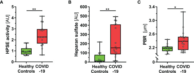
Figure 1.
COVID-19 patients show elevated HPSE activity, and damaged eGC in vivo. (A-C) Boxplots showing (A) heparanase (HPSE) activity, (B) heparan sulfate (HS) and (C) perfused boundary region (PBR; an inverse estimate of the sublingual endothelial glycocalyx thickness) in healthy subjects (n = 12) and COVID-19 patients at the ICU (n = 16). Differences between groups were calculated by Mann-Whitney U test. *p < 0.05; **p < 0.001.

