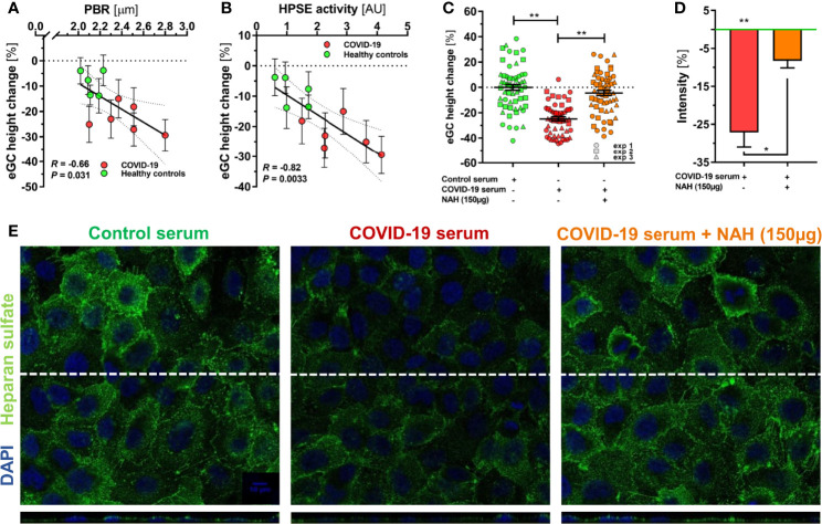
Figure 2.
HPSE is a putative mediator of eGC damage in COVID-19. (A-C) Sera from a randomly selected subgroup of 5 healthy controls and 6 COVID-19 patients were sterile-filtered and incubated (5%) on the human umbilical vein endothelial cell line EA.hy926 for 60 min. Endothelial glycocalyx (eGC) thickness was assessed by atomic force microscopy (AFM) using a dedicated nano-indentation protocol. Scatter dot plot showing the association between AFM-derived eGC (in vitro) decline and corresponding (A) PBR-values (in vivo) and (B) HPSE activity for the individuals from the subgroup. Each dot represents the mean ± SEM (standard error of mean) of two independent AFM experiments (consisting of ≥ 4 indentation curves in each of ≥ 8 different cells) for each individual serum. Incubation without human serum served as control. Correlation was assessed by Spearman correlation coefficient. (C) Dot plots from three independent AFM experiments (pooled serum from subgroups) showing values with mean ± SEM. Each dot represents the mean of ≥ 4 indentation curves per cell. Heparanase was blocked by N-desulfated re-N-acetylated heparin (NAH; 150 µg). Differences between groups were calculated with nested ANOVA and Tukey’s post-hoc test. Intensity analysis of heparan sulfate-stained EA.hy926 cells (D) and representative immunofluorescence images (E) after treatment with 5% control serum or COVID-19 serum ± NAH (150 µg) for 60 min. Values are normalized to control serum (zero line) and differences between groups were assessed with nested ANOVA and Tukey’s post-hoc test. Data are presented as mean ± SEM. *p < 0.05; **p < 0.001.

