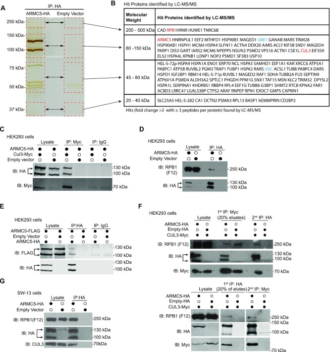
Figure 1.
ARMC5 forms a complex with CUL3, RPB1, and itself. (A) Silver staining of ARMC5 precipitates resolved by SDS-PAGE. Lysates of HEK293 cells transfected with ARMC5-HA-expressing constructs were precipitated with anti-HA Ab. The precipitates were resolved by SDS-PAGE. The regions (rectangles with dashed lines) with visible bands in the test sample and the corresponding positions in the empty vector-transfected lane were excised and were analyzed by LC–MS/MS. Three independent experiments were conducted, and a representative gel with silver staining is shown. (B) Proteins found in the ARMC5 precipitates according to the LC–MS/MS analysis. Proteins met with both the following two conditions in any of the biological replicates (200-500 kDa: duplicates; 80–150 kDa: duplicates; 45–80 kDa: triplicates; 30 kDa: no replicate) were listed. 1) The protein had equal or more than three peptides corresponding to its sequence in the ARMC5-HA transfected sample; 2) the number of the peptides in the ARMC5-HA-transfected sample was more than 2-fold larger than that in the empty vector control. The gel pieces from which the proteins were derived were indicated. (C) ARMC5 interacted with CUL3. HEK293 cells were transfected with plasmids expressing ARMC5-HA and CUL3-Myc. Cell lysates were precipitated with anti-HA Ab and immunoblotted with anti-Myc Ab. (D) ARMC5 interacted with RPB1. HEK293 cells were transfected with plasmids expressing ARMC5-HA. Cell lysates were precipitated with anti-HA Ab and immunoblotted with anti-RPB1 N-terminal Ab (clone F12). (E) ARMC5 interacted with itself. HEK293 cells were transfected plasmids expressing ARMC5-HA and ARMC5-FLAG. Cell lysates were precipitated with anti-HA Ab and immunoblotted with anti-FLAG Ab. (F) ARMC5, CUL3, and RPB1 formed tri-molecule complexes. HEK293 cells were transfected with plasmids expressing ARMC5-HA and CUL3-Myc. In the upper panel, cell lysates were first precipitated with anti-Myc Ab and eluted with Myc peptides. The precipitates were then re-precipitated with anti-HA Ab. The secondary precipitates were blotted with anti-RPB1 N-terminus Ab (clone F12). In the lower panel, the order of precipitation was reversed. The lysates were first precipitated with anti-HA Ab. The precipitates were then precipitated with anti-Myc Ab. (G) ARMC5 interacted with endogenous CUL3 and RPB1 in adrenal cortical carcinoma SW-13 cells. SW-13 cells were transfected with plasmids expressing ARMC5-HA. Cell lysates were precipitated with anti-HA Ab. The precipitates were immunoblotted with anti-CUL3 Ab or anti-RPB1 N-terminus Ab (clone F12). In all the experiments, the lysates were also immunoblotted with Abs against HA, MYC, or FLAG, as applicable, to demonstrate the effectiveness of transfection. Empty vectors were used in transfection as controls. IgG was employed in immunoprecipitation as a control. All the experiments were conducted more than three times, and representative results are shown.

