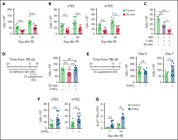
Figure 2.
Dietary Zn intake dictates regenerative capacity of the thymus after damage. (A-B) Six- to 8-week-old female C57BL/6 mice were fed a normal or ZD diet for 21 days, at which point mice were given a sublethal dose of TBI (550 cGy). (A) Total thymic cellularity at days 7 and 14 after TBI (n = 10/treatment per timepoint across 2 independent experiments). (B) Total number of CD45−EpCAM+MHCII+UEA1loLy51hi (cTECs) and CD45−EpCAM+MHCII+UEA1hiLy51lo (mTECs) at days 7 and 14 after TBI (n = 10/treatment per timepoint across 2 independent experiments). (C) Six- to 12-week-old female BALB.B mice were fed with normal or ZD diet for 21 days, at which point mice were given a lethal dose of TBI (900 cGy) and 10 × 106 T-cell–depleted BM cells from 6- to 8-week-old C57BL/6 mice. One cohort also received 2 × 10⁶ T cells to induce GVHD; thymic cellularity was quantified on day 14 after allo-HCT (n = 5-6/group). (D) Six- to 8-week-old female C57BL/6 mice were fed with normal or ZD diet for 21 days, at which point mice were given 550 cGy TBI and total thymus cellularity quantified on day 14. One cohort was given supplemental Zn in drinking water (300 mg/kg per day of ZnSO4) from day 0 until analysis on day 14 (n = 10/group across 2 independent experiments). (E-F) Six- to 8-week-old female C57BL/6 mice were given supplemental Zn in drinking water (300 mg/kg per day ZnSO4) for 21 days, at which point mice were given 550 cGy of TBI. Mice were maintained on ZnSO4 in drinking water and the thymus was analyzed on day 0 or 7 (day 0: untreated, n = 22; ZD, n = 10 across 2 independent experiments; day 7: untreated, n = 15; ZD, n = 15, across 3 independent experiments). (E) Total thymic cellularity. (F) Total number of cTECs and mTECs. (G) Total number of Ki-67+ TECs. Graphs represent mean ± SEM; each dot represents a biologically independent observation. *P < .05; **P < .01; ***P < .001.

