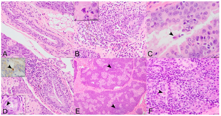
Figure 2.
Histopathologic lesions in the salivary glands of SOSV-infected Egyptian rousette bats euthanized at 6, 9 and 21 DPI. (A) Salivary gland, control bat 290195. A normal interlobular duct is 1–3 cell layers thick and contains very low numbers of intraepithelial and peri-ductular resident immune cells. HE stain; original magnification = 400×; bar = 20 µm. (B) Salivary gland, 6 DPI bat 284899. Acute sialodochitis characterized by a robust infiltrate of mononuclear and granulocytic inflammatory cells that expands the duct wall and surrounding connective tissue. Granulocytic cells, mononuclear cells and cellular debris fill and partially occlude the duct lumen. Inset: The luminal aspect of a different, but similarly affected duct contains an epithelial syncytium and several granulocytes. HE stains; original magnifications = 400× (main image; bar = 20 µm) and 1000× (inset; bar = 10 µm). This bat was qRT-PCR positive for SOSV in the salivary gland at necropsy and had SOSV RNA in the oral swabs on days 5 and 6 PI. (C) Salivary gland, 9 DPI bat 287014. The duct is multifocally infiltrated by low numbers of mononuclear and granulocytic cells. The lumen contains low numbers of neutrophils and a binucleated cell with abundant granular cytoplasm (suspected syncytium) (arrowhead). HE stain; original magnification = 400×; bar = 20 µm. (D) Salivary gland, 21 DPI bat 289338. An interlobular duct is focally infiltrated and expanded by a mixture of mononuclear and granulocytic inflammatory cells. Bottom left inset: The wall of a smaller but similarly affected duct contains an apoptotic body that is shrunken and rounded with distinct cell borders and a pyknotic nucleus (arrowhead). HE stains; original magnifications = 400× (main image; bar = 20 µm) and 1000× (inset; bar = 10 µm). Top left inset: An apoptotic body in the wall of the same duct as the lower left inset has diffuse, moderate cytoplasmic immunoreactivity for cleaved caspase 3 (arrowhead). Immunohistochemical stain with 3-3′-Diaminobenzidine (DAB; brown) chromogen to visualize cleaved caspase 3 antigen and hematoxylin counterstain. Original magnification = 600×; bar = 20 µm. (E) Salivary gland, 21 DPI bat 290040. Low magnification image of salivary gland acinar tissue showing multiple foci of mononuclear cells which expand the interstitium and focally obscure the acini (arrowhead). HE stain; original magnification = 200×; bar = 50 µm. (F) Salivary gland, same 21 DPI bat as previous image. Higher magnification of one hypercellular focus showing interstitial expansion with numerous mononuclear inflammatory cells. One intralobular duct epithelial cell exhibits cytoplasmic swelling, peripheralization of nuclear material and accumulation of eosinophilic material within the nucleus suggestive of intranuclear inclusion material (arrowhead). HE stain; original magnification = 400×; bar = 20 µm.

