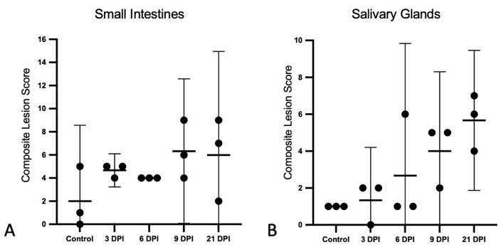
Figure 3.
Histopathologic lesion scores in the small intestines and salivary glands of SOSV-infected Egyptian rousette bats. Individual groups are represented on the x axis and each dot represents the score for a single bat. Bars represent the mean and lines represent the ±95% confidence interval. Control bats were euthanized at 21 DPI. (A) Criteria used to generate a composite small intestine lesion score for each bat included: villus epithelial discontinuity or ulceration, accumulation of eosinophilic fluid in the lamina propria, hypercellularity of the lamina propria, epithelial cell apoptosis and/or tingible body macrophages in the lamina propria and villus fusion. (B) Salivary gland scores were positively correlated with time. Criteria used to generate a composite salivary gland lesion score for each bat included: sialodochitis, sialadenitis, duct epithelial cell apoptosis and epithelial syncytia.

