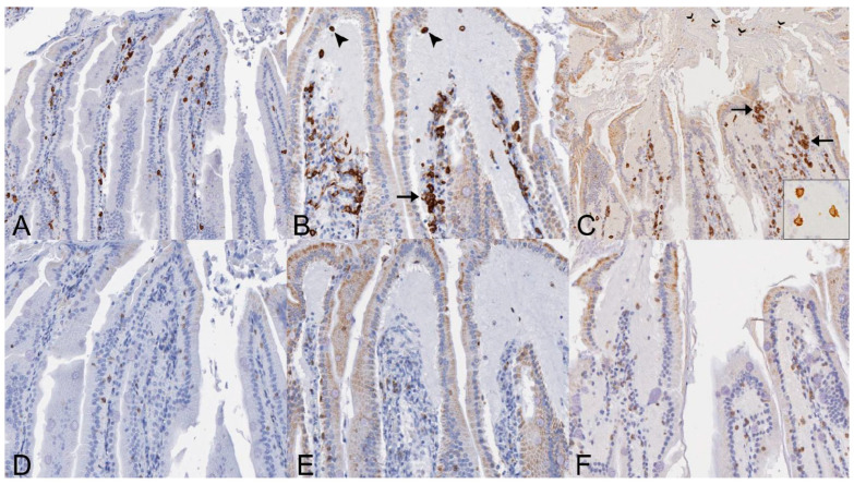
Figure 6.
Distribution of Iba1- and CD3-immunolabeled cells in the small intestines of SOSV-infected Egyptian rousette bats euthanized at 9 and 21 DPI. (A) Iba1 immunohistochemistry (IHC), jejunum, control bat 290195. Iba1-immunolabeled cells (brown) are present in moderate numbers throughout the lamina propria core of the small intestinal villi. Original magnification = 200×. (B) Iba1 IHC, jejunum, 9 DPI bat 287756. Moderate numbers of Iba1-immunolabeled cells are present within the lamina propria core, occasionally in small clusters (arrow). Low numbers of Iba1+ cells with an amoeboid morphology are scattered within the lamina propria fluid and just beneath the epithelium at the villus tip (arrowheads). The small intestines of this bat had the highest viral load of all tissues tested during the serial sacrifice study. Original magnification = 200×. (C) Iba1 IHC, jejunum, 21 DPI bat 289953. The villus core and lamina propria contain moderate numbers of Iba1+ cells, occasionally clumped in small aggregates (arrows). Several villus tips have epithelial defects through which fluid spills from the lamina propria into the lumen. This luminal fluid contains several Iba1+ cells (open arrowheads). Inset: Higher magnification image demonstrating the amoeboid phenotype of macrophages present within the lamina propria fluid. This bat intermittently shed SOSV in the feces throughout the duration of the serial euthanasia study. Original magnifications = 100× (main image) and 600× (inset). (D) CD3 IHC, jejunum, control bat 290195. Low numbers of CD3+ cells (brown) are scattered throughout the epithelium. Rare CD3+ cells are present within the lamina propria core. Original magnification = 400×. (E) CD3 IHC, jejunum, 9 DPI bat 287756. Low numbers of CD3+ cells are scattered throughout the intestinal epithelium and occasionally present within the lamina propria fluid. Slightly increased CD3+ cells are present within the lamina propria core compared to the control. Original magnification = 400×. (F) CD3 IHC, jejunum, 21 DPI bat 289953. Scattered CD3+ cells are present just beneath the intestinal epithelium and within the lamina propria fluid. Slightly increased numbers of CD3+ cells are distributed throughout the lamina propria core compared to the control. Original magnification = 400×. All immunohistochemical stains were performed using 3-3′-Diaminobenzidine (DAB; brown) chromogen to visualize cellular antigen and hematoxylin counterstain.

