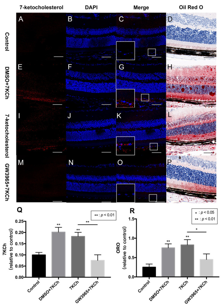
Figure 2.
7KCh immunofluorescence and Oil Red O staining of retinas. 7KCh deposition was detected by immunofluorescence (A–C,E–G,I–K,M–O) and Oil Red O staining (D,H,L,P). (A–D) C57BL/6 mice were gavaged saline (100 μL/10 g) for seven days (n = 4). (E–H) Firstly, C57BL/6 mice were gavaged DMSO (1%) for three days, and then mice were subretinally injected 7KCh. After injection, gavaging with DMSO (1%) continued for four days. (I–L) C57BL/6 mice were gavaged saline (100 μL/10 g) for three days, then subretinally injected 7KCh. After injection, gavaging with saline (100 μL/10 g) continued for four days. (M–P) C57BL/6 mice were gavaged GW3965 (10 mg/kg) for three days, then the mice were subretinally injected 7KCh. After injection, gavaging with GW3965 (10 mg/kg) continued for four days. Retinas were harvested, sectioned, and subjected to double immunofluorescent staining and Oil Red O staining analyses. Scale bar 50 μm. (Q,R) 7KCh immunofluorescence and Oil Red O intensity were averaged at eight locations. Data are represented as mean ± SEM of three independent experiments. Differences of p < 0.05 were considered statistically significant.

