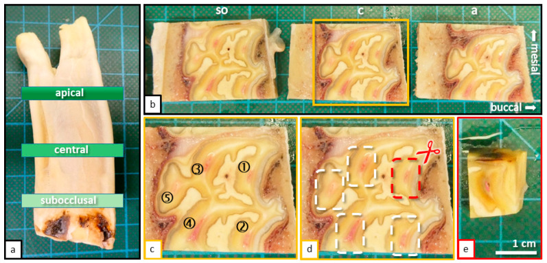
Figure 2.
(a) Cheek tooth of an adult horse (208), sectioned horizontal levels (subocclusal, central, and apical) are indicated. (b) Sections of the tooth (208) displayed in Figure 1a, occlusal view, sectioned with a diamond coated micro-band saw. Horizontal sections were taken from three defined levels: Subocclusal (so); Central (c); Apical (a). (c) Magnification of the central horizontal section with indicated pulp horns (1–5). (d) Same image as shown in Figure 1b. The process of further sectioning and separation of pulp horns is displayed. (e) Final specimen before placement in embedding cassettes.

