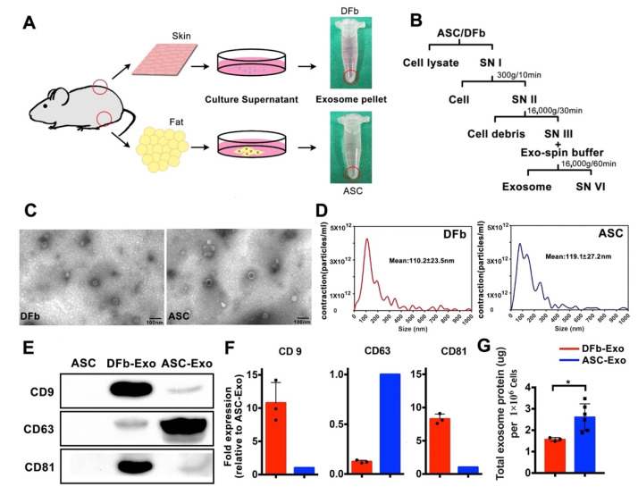
Figure 1.
Characterization of dermal fibroblast (DFb) and adipose stem cell (ASC) derived exosomes. (A) Schematic illustrating the workflow used to generate exosomes. The arrows indicate harvested dermal fibroblast or adipose stem cell-derived exosome pellets; (B) differential centrifugation procedure for the isolation of exosomes from DFb and ASC culture supernatants (SN); (C) transmission electron microscopic morphological analysis of ASC-Exo and DFb-Exo depicts multiple cup-shaped and shrunken vesicles in the conditioned medium (scale bar: 100 nm); (D) size distribution of ASC-Exo and DFb-Exo based on NanoSight nanoparticle tracking analysis (NTA). Statistics of the size of ASC-Exo and DFb-Exo based on NTA are shown as mean ± SD (representative result of four independent analyses); (E) Western blot analysis of general exosome markers, including CD9, CD63, and CD81 for ASC-Exo and DFb-Exo; (F) semi-quantification analysis of CD63, CD9, and CD81 expression from ASC-Exo and DFb-Exo; (G) total protein contained within exosomes isolated from the culture supernatant of 1 × 10⁶ dermal fibroblasts (n = 3) and adipose stem cells (n = 6). * p < 0.05. Error bars are mean ± SE. Data were analyzed using independent unpaired two-tailed Student’s t-tests.

