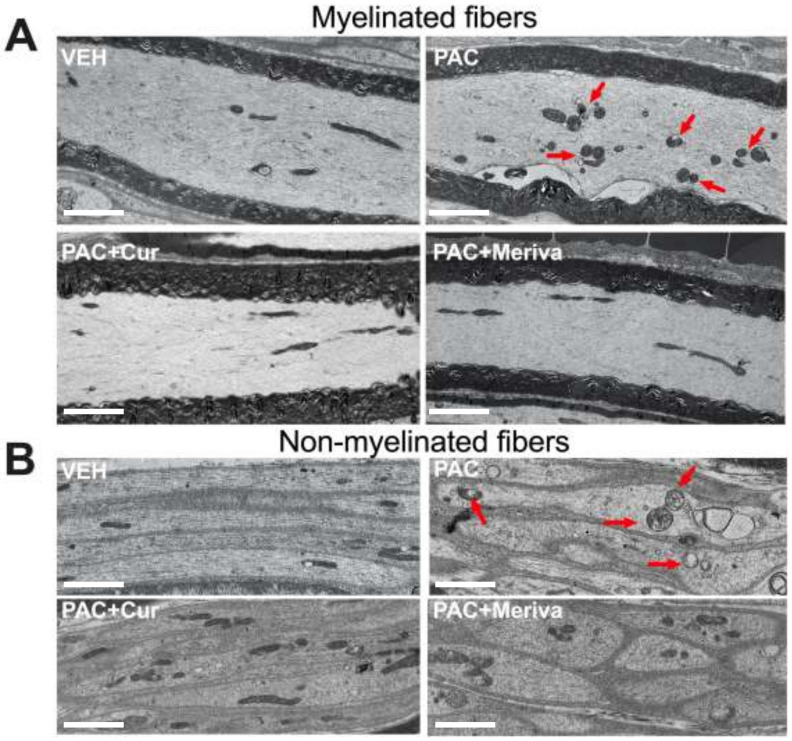
Figure 3.
Morphological features of sciatic nerves: transmission-electron-microscopy pictures of sciatic-nerve longitudinal sections. Pictures showing mitochondrial ultrastructure in the control group (VEH), the paclitaxel-treated group (PAC), the paclitaxel + curcumin 1.5%-treated group (PAC + Cur) and the paclitaxel + Meriva 1.5%-treated group (PAC + Meriva), in myelinated (A) and non-myelinated fibers (B). The red arrows show mitochondria with a pathological aspect. Bar scales: 2.5 μm.

