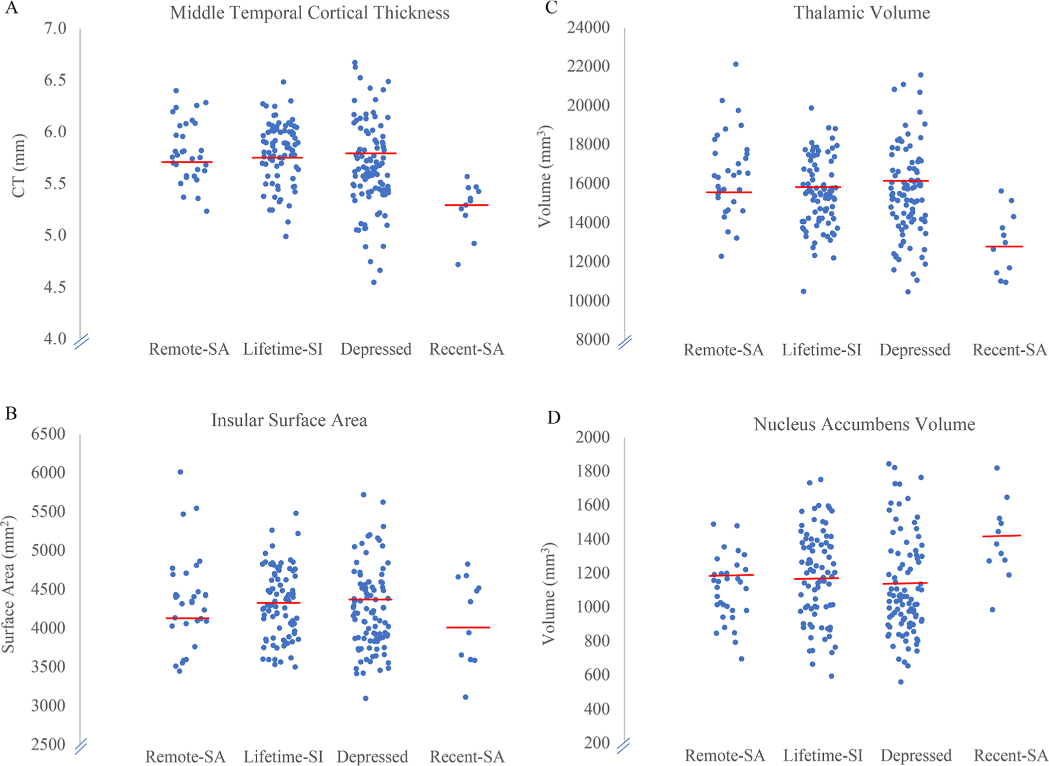
Fig. 1.

Scatterplots of cortical thickness, surface area, and volume in the 4 groups. A) Lower middle temporal cortical thickness was observed in the Recent-SA and Lifetime-SI only groups as compared to the non-suicidal depressed group. B) Lower insular surface area was observed in the Recent-SA group as compared to the Lifetime-SI only and non-suicidal depressed groups. C) Lower thalamic volume was observed in the Recent-SA group as compared to the Lifetime-SI only and non-suicidal depressed groups. D) Higher volume in the nucleus accumbens was observed in the Recent-SA group as compared to the Lifetime-SI only and non-suicidal depressed groups.
