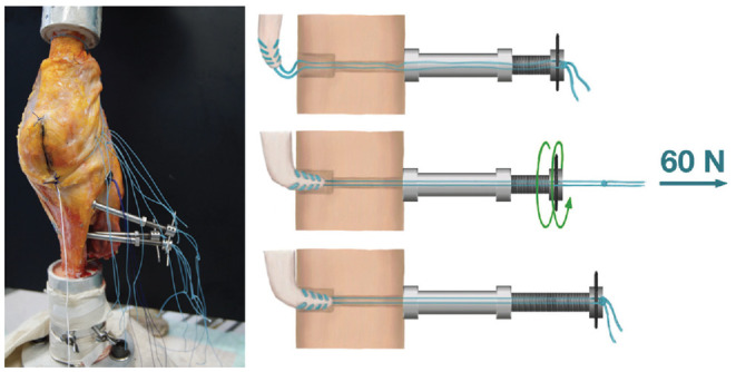
Figure 3.

With the knee in upright position (femur on top, tibia at the bottom), custom-made tensioning devices were installed at the opposite cortex to allow for separate tensioning of each medial reconstruction.

With the knee in upright position (femur on top, tibia at the bottom), custom-made tensioning devices were installed at the opposite cortex to allow for separate tensioning of each medial reconstruction.