
Figure 3. Lipid-coated EuCeO2NPs facilitate BV2 phagocytosis visualized by transmission electron microscopy (TEM).
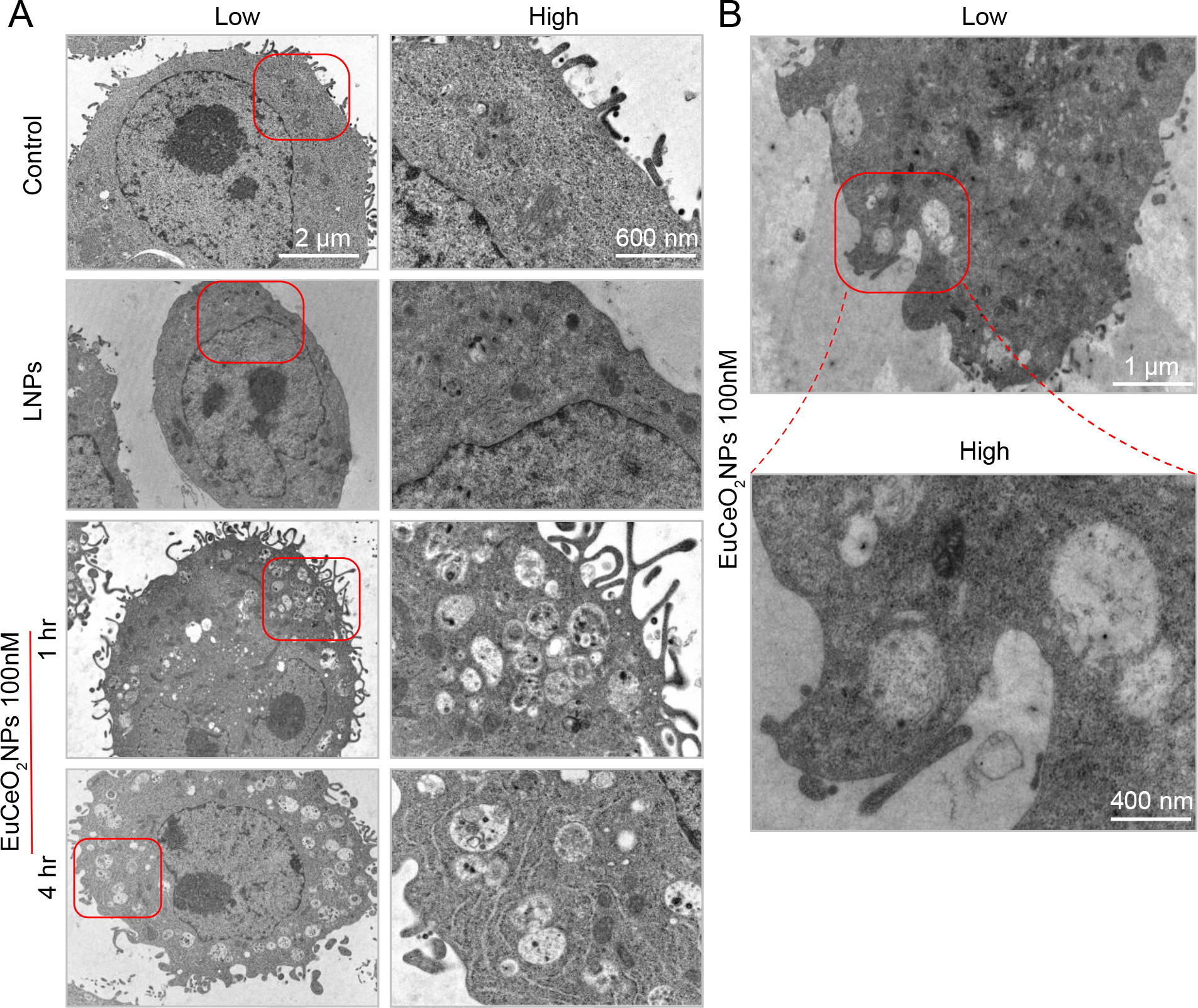
(A) Low and high magnification images of fixed BV2 cells from either untreated (control) or 100 ng/ml LNPs or EuCeO2NPs treated cells were observed for nanoparticle uptake after 1 and 4 hr of treatment. (B) EuCeO2NPs treated BV2 cells demonstrated nanoparticle phagocytosis following 1hr incubation.