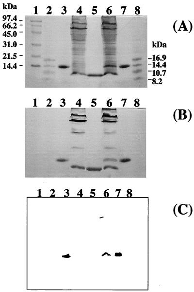
FIG. 1.
SDS-PAGE analyses of cytochrome c3 in S. oneidensis. Cell preparations were loaded onto SDS–15% PAGE gels and electrophoresed. (A) Protein staining with CBB; (B) heme staining with o-tolidine dihydrochloride heme stain prepared as previously described (2); and (C) antigenically active material via Western blotting with antibody to cytochrome c3 from D. vulgaris Miyazaki F. Lanes 3, 5, and 7 were loaded with ca. 0.005 mg of protein, and lanes 4 and 6 were loaded with ca. 1 mg of protein. Lane 1, high-molecular-mass markers (phosphorylase b [97.4 kDa], bovine serum albumin [66.2 kDa], ovalbumin [45 kDa], carbonic anhydrase [31 kDa], soybean trypsin inhibitor [21.5 kDa], and lysozyme [14 kDa]); lanes 2 and 8, low-molecular-mass markers (globin [16.95 kDa], globins I and II [14.4 kDa], globins I and III [10.7 kDa], and globin I [8.16 kDa]); lanes 3 and 7, wild-type cytochrome c3 from D. vulgaris Miyazaki F; lane 4, cell lysate from S. oneidensis MR-1; lane 5, wild-type cytochrome c3 from S. oneidensis MR-1; lane 6, cell lysate from S. oneidensis TSP-C(pRKMα).

