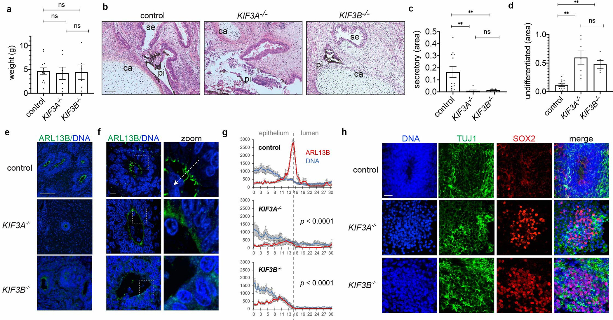
Fig. 3. Cilia are critical for terminal differentiation in vivo.

(a) Weights of growths removed from animals (mean ± s.e.m., n ≥ 6 independent biological replicates per condition from a total of 14 distinct cell lines). (b) Representative images and (c-d) quantification of tissue types in growth sections stained with hematoxylin and eosin. Labels indicate pigmented epithelium (pi), secretory epithelium (se), and cartilage (ca) regions. (mean ± s.e.m., n ≥ 5 independent biological replicates per condition from a total of 14 distinct cell lines; **, p < 0.01.) (e) Representative wide-field and (f) confocal microscope images showing ARL13B, with (g) averaged line scans drawn from neuroepithelium into rosette lumens (mean ± s.e.m., n ≥ 9 independent biological replicates per condition from a total of 8 distinct cell lines). White dashed arrow illustrates how line scans were drawn. (h) Representative confocal images showing neuronal marker immunofluorescence in teratomas. Scale bar, 100 μm (b,e) or 10 μm (f,h).
