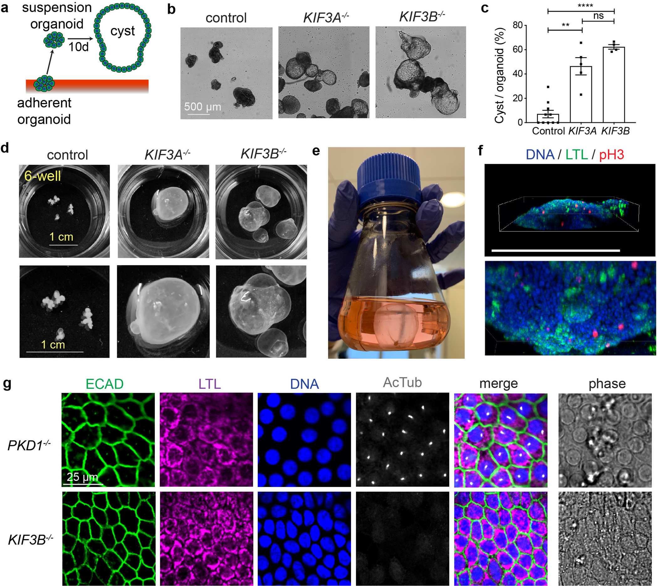
Fig. 7. Kinesin-2 knockout organoids express the PKD cystic phenotype.

(a) Schematic of cystogenesis assay. (b) Representative phase-contrast images of control, KIF3A−/−, and KIF3B−/− organoids after 10 days in suspension culture. Scale bars, 500 μm. (c) Percentage of cystic organoids in suspension cultures (mean ± s.e.m., n ≥ 4 biological replicates per condition from a total of ten distinct cell lines. **, p < 0.01; ****, p < 0.0001). (d) Representative photographs of whole 6-wells containing organoids grown for several months in suspension culture, with zoom below. Scale bars, 1 cm. (e) 250 ml flask containing a large KIF3B−/− cystic organoid that outgrew the 6-well plates, as shown in Movie 1. (f) Scenes from Movie 2 showing 3-D confocal reconstruction of the lower edge of a cyst labeled with phospho-histone H3 (pH3) for mitotic cells and LTL for proximal tubular epithelia. Scale bar, 500 μm. (g) Confocal immunofluorescence of cilia (AcTub) and kidney epithelial cell markers in cyst-lining epithelial cells. Scale bar, 25 μm.
