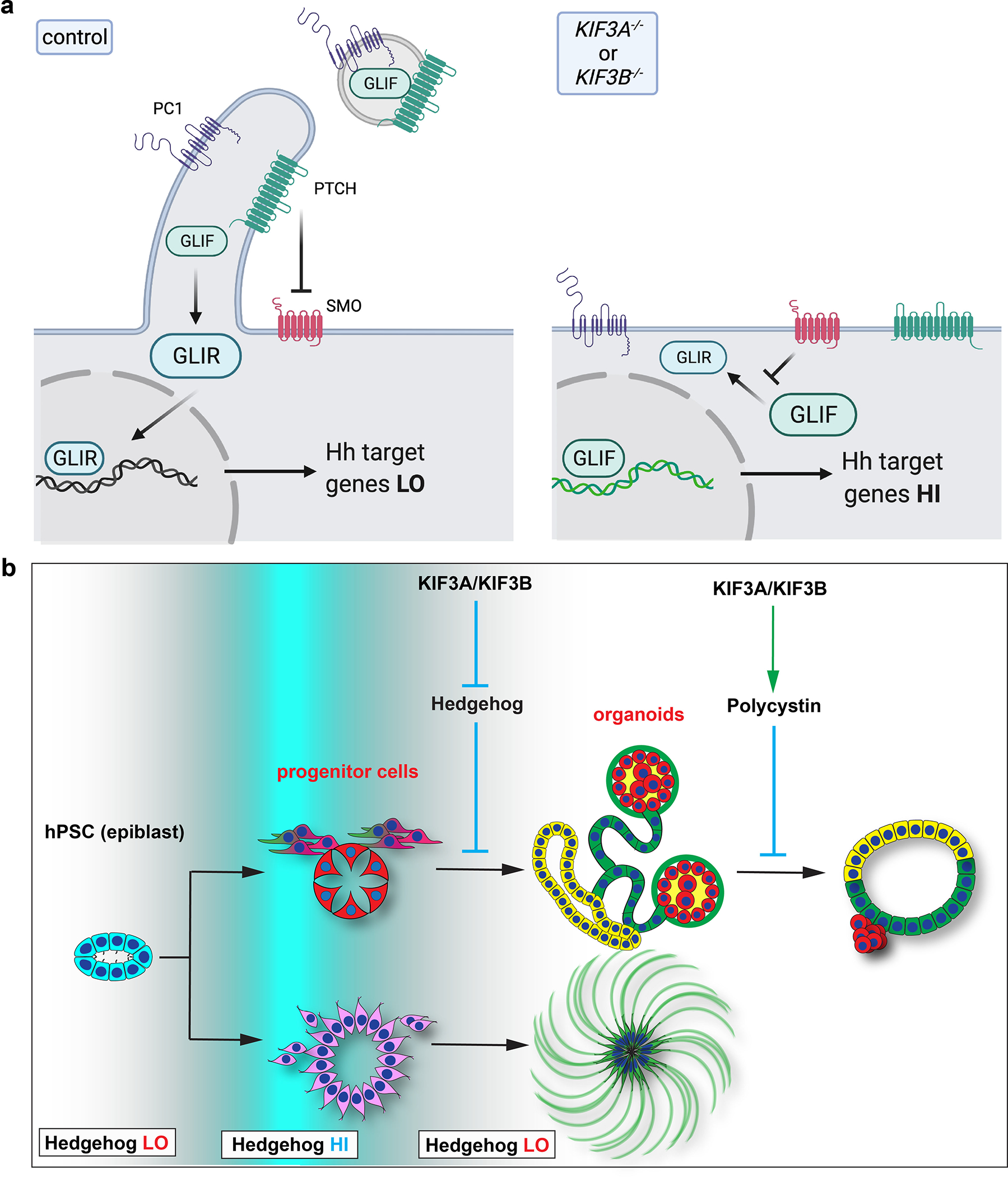
Fig. 9. Ciliary function directs morphogenesis in complex tissues derived from hPSCs.

(a) Schematic of kinesin-2’s effects at the cell surface. Left: In normal cells, the cilium generated by kinesin-2 is inhabited by PTCH1 which establishes a protected zone to promote downstream GLI protein processing (relative quantity of GLIF versus GLIR), ultimately resulting in proper modulation of the hedgehog signal and levels of downstream gene expression. Hedgehog and PKD components exit the cell through ciliary EV to further modulate signaling. Right: In kinesin-2 knockout cells, absence of the cilium renders PTCH1 unable to inhibit SMO, resulting in blockade of GLI protein processing and increased activation of the hedgehog signaling pathway. Hedgehog and PKD components are unable to exit the cell. (b) Illustration depicting the role of cilia in tissues and organoids derived from hPSCs. Kinesin-2 activity is responsible for switching the hedgehog signaling between ‘HI’ and ‘LO’ states (white to blue gradient) during differentiation of the pluripotent epiblast into complex tissues (left to right). In differentiated tubular epithelia, kinesin-2 regulates PKD pathway components to restrict expansion into cysts.
