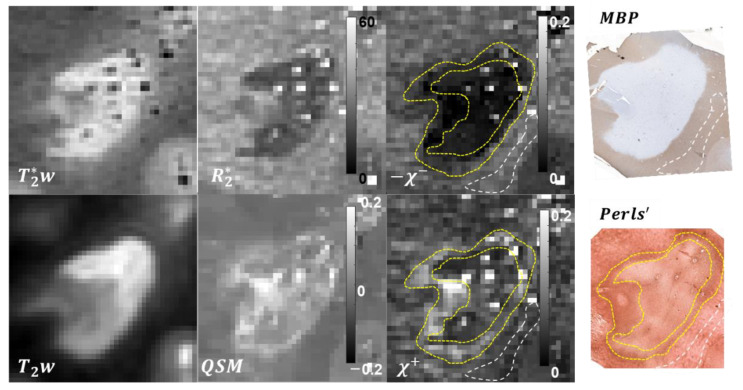
Figure 1.
Results of the -based separation of magnetic sources in a chronic active lesion. Paramagnetic lesion rim readily identifiable in QSM and (yellow dashed line) appears to be in good morphological agreement with the iron distribution revealed by Perls’ staining. Similarly, strong demyelination of the lesion core estimated with the proposed method is well reflected by the MBP staining. NAWM is shown with white dashed line.

